
Pancreatitis
Introduction
|
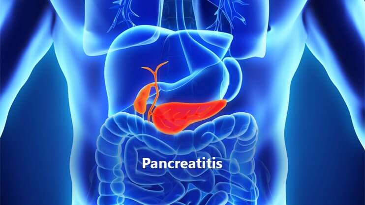
Pancreatitis is a syndrome characterized by inflammation of the pancreas. The pancreas is a long, flat gland that sits tucked behind the stomach in the upper abdomen. The pancreas produces digestive enzymes and a number of hormones that help regulate the way the body processes sugar (glucose). There are two types: acute pancreatitis and chronic pancreatitis. Both are discussed in this literature. |
Acute Pancreatitis
The twin challenges of acute pancreatitis are to establish the diagnosis and stratify severity. The difficulty in diagnosing pancreatitis lies in its non-specific symptomatology, which is shared by a number of other gastrointestinal diseases.
Patient outcome may be dependent on prompt recognition of severe pancreatitis. These patients require aggressive treatment to reverse organ failure and admission to intensive care or a high-dependency area for ongoing management. The hunt for the aetiology is the next priority, but this may be deferred to the inpatient team.
1. Pathogenesis and Aetiology
The pathogenesis of acute pancreatitis relates to inappropriate activation of trypsinogen to trypsin, which in turn releases digestive enzymes causing pancreatic injury. In 20% of cases, when pancreatic necrosis occurs it is coupled with infection from translocation of gut bacteria. An inflammatory response ensues, resulting in systemic inflammatory response syndrome, multiorgan dysfunction syndrome and, in some cases, death.
The commonest risk factor for pancreatitis in males is excessive alcohol use, and in females gallstone disease. The other aetiological factors are listed below (Table X1).
| Table X1 | Aetiologes of Acute Pancreatitis |
|---|
CommonGallstone (including microlithiasis)Alcohol Idiopathic Dyslipidaemia Hypercalcaemia (hyperparathyroidism, metastatic bone disease, sarcoidosis) Sphincter of Oddi dysfunction Drugs (azathioprine, valproate, pentamidine, didanosine, co-trimoxazole) Toxins Post ERCP Traumatic Post operative |
UncommonStructural (cancer of the pancreas/periampullary, pancrease divisum)Vasculitis |
RareInfective (Coxsackie, mumps, HIV, parasitic, ascariasis)Autoimmune (systemic lupus erythematosus, Sjögren’s syndrome) α1-Antitrypsin deficiency |
2. Epidemiology
The incidence of pancreatitis is rising, reflecting an increase in alcohol consumption and gallstone disease. However, despite advances in care, overall mortality remains unaltered at 2–10%.
3. Clinical Features
Gallstone pancreatitis typically presents with a sudden onset of severe, constant epigastric pain radiating to the back. In contrast, pain in pancreatitis from other causes (e.g., alcohol) has a more insidious onset and may be poorly localized. This is often accompanied by nausea and vomiting. Abdominal wall ecchymosis around the umbilicus (Cullen’s sign), flanks (Grey Turner’s sign) and inguinal ligament (Fox’s sign — an uncommon finding) does not occur till 36–72 hours after the onset of pain.
4. Differential Diagnosis
The most important differential diagnoses to exclude are perforated viscus, ischaemic colitis, leaking abdominal aortic aneurysm and myocardial ischaemia.
5. Clinical Investigations
Amylase rises in 2–12 hours and normalizes in about a week. In 10% of cases of pancreatitis, amylase is falsely negative due to depleted acinar cell mass. False positives may occur with salivary gland disease, macroamylasaemia and some cancers.
Lipase rises in 4–8 hours and normalizes in 1–2 weeks. It has superior sensitivity and specificity compared to amylase, as it is only produced in the pancreas. Amylase or lipase levels more than three times the upper limit of normal are diagnostic of acute pancreatitis.
Lesser elevations must be interpreted against the timing of the test from symptom onset. The peak amylase and/or lipase level does no correlate with the severity of the disease.
When clinical signs and biochemical tests are equivocal, a contrast-enhanced CT scan of the abdomen is the radiological investigation of choice as it can establish the diagnosis, exclude most of the differential diagnoses listed above, stage the disease and detect complications. The use of ultrasound is not as helpful as the pancreas is poorly seen in 25–50% of patients, though it may show gallstones and/or a dilated common bile duct, giving a clue to its aetiology.
Plain radiography of the chest and abdomen has poor sensitivity for the diagnosis. Chest X-ray may shoe a pleural effusion or features of acute respiratory distress syndrome, and abdominal films may show gallstones, a sentinel bowel loop or peripancreatic retroperitoneal gas, the latter signifying infection of the pancreas.
Other tests to aid severity scoring and identification of aetiology include full blood count with haematocrit, urea, electrolytes, lactate dehydrogenase, alanine aminotransferase, blood gas analysis, calcium and lipid profile.
6. Severity Scoring
6.1 Biochemical
Severe pancreatitis is identified either using a predictive scoring system or when a patient presents in frank organ failure (e.g. respiratory, renal or cardiovascular). Of the three predictive severity scoring systems in use, only the APACHE II allows scoring at presentation. A score ≥8 indicates severe pancreatitis with a mortality of 11-18%. The Ransom and Glasgow scores (Table X2) can only be completed at 48 hours, which limits their usefulness in the ED. Likewise, an elevated C-reactive protein (CRP) > 150 mg/dL 24 or 48 hours after presentation also reliably predicts severe pancreatitis.
| Table X2 | Biochemical severity scoring systems | |
|---|---|
| Ranson’s score (1 point for each positive factor, Score ≥3 indicates severe pancreatitis) | |
At presentation | |
| Age | > 55 yr |
| Blood glucose | > 10 mmol/L |
| White cell count | > 16 000/mm3 |
| Lactate dehydrogenase | > 350 IU/L |
| Alanine aminotransferase | > 250 IU/L |
Within 48 h after presentation | |
| Haematocrit | > 10% decrease |
| Calcium | < 2 mmol/L |
| Base deficit | > 4 mEq/L |
| Urea | > 1.8 mmol/L increase since admission |
| Fluid sequestration | > 6 L |
| Partial pressure of arterial oxygen | < 60 mmHg |
Glasgow scoring system for prediction of severity in acute pancreatitis (1 point for each positive factor. Score ≥3 indicates severe pancreatitis) | |
| Partial pressure of arterial oxygen | < 60 mmHg |
| Albumin | < 32 g/L |
| Calcium | < 2 mmol/L |
| White cell count | > 15 000/mm3 |
| Aspartate aminotransferase | > 200 IU/L |
| Lactate dehydrogenase | > 600 IU/L |
| Blood glucose | > 10 mmol/L |
| Urea | > 16 mmol/L |
6.2 Radiological
Once severe pancreatitis is identified, contrast-enhanced CT is used to anatomically score the severity using the system described by Balthazar.
In summary, severe pancretitis should be considered at presentation if the following risk factors are present: age>65 years, body mass index >30 kg/m2, presence of pleural effusion on chest X-ray, contrast-enhanced CT shows >30% necrosis, APACHE II score ≥8, symptoms and signs of organ failure (e.g. poor urine output, progressive tachycardia, tachypnoea, hypoxaemia, agitation, confusion, rising haematocrit level).
7. Emergency Department Treatment
The treatment for acute pancreatitis is supportive, with emphasis on fluid replacement and prevention of hypoxia.
- Supplemental oxygen. Hypoxia may indicate ARDS or significant pleural effusions. Mechanical ventilation may be required in patients with respiratory distress. Uncorrected, gut hypoxia promotes translocation of Gram-negative bacteria.
- Fluid resuscitation. Significant third-space losses may occur. Fluid replacement should be titrated to blood pressure and urine output. A worsening haematocrit indicates insufficient replacement. Central venous monitoring should be considered in severe cases.
- Analgesia. Opioid analgesia is often required. Morphine, administered intravenously, is the agent of choice and dose should be titrated against response. On occasion, large doses are required for pain control. There are no human studies to support the belief that morphine causes spasm of the sphincter of Oddi.
- Disposition. Patients with pancreatitis require admission for treatment and observation of disease progression. Mild pancreatitis can be managed in the general ward, but severe pancreatitis should be managed in intensive care or a high-dependency unit.
8. Prognosis
The majority of patients with acute pancreatitis experience a mild, self-limiting course. Twenty per cent of patients develop severe pancreatitis, with a mortality of 20%; 50% of deaths occur in the first week from multiorgan dysfunction syndrome, whereas death after 1 week is usually due to infective complications. If organ failure is reversed within 48 hours, the prognosis is good.
9. Complications
Local complications include pancreatic pseudocyst, abscess, splenic vein thrombosis, duodenal obstruction and progression to chronic pancreatitis. Systemic complications include hypocalcaemia, pleural effusion, ARDS and multiorgan dysfunction syndrome.
Chronic Pancreatitis
Patients with chronic pancreatitis may present with recurrent abdominal pain radiating to the back. This may be associated with weight loss because of fear of eating due to postprandial exacerbations of pain. There may be signs of pancreatic exocrine insufficiency (steatorrhoea) or endocrine insufficiency (diabetes mellitus).
Physical examination may reveal a mass in the epigastrium, suggesting a pseudocyst, and the patient may assume a characteristic pain-relieving posture of lying on the side with the knees drawn up to the chest.
The aetiology of chronic pancreatitis is usually metabolic in nature, with excessive alcohol consumption accounting for 60–90% of cases. The primary process is chronic irreversible inflammation, fibrosis and calcification of the pancreas, affecting its exocrine and endocrine functions.
In chronic pancreatitis, serum amylase and lipase levels are not as elevated as in acute pancreatitis. Occasionally enzyme levels may be normal due to atrophy of the gland. ERCP is the gold standard for diagnosis of chronic pancreatitis. Contrast-enhanced CT and magnetic resonance cholangiopancreatography are non-invasive and provide information about the pancreatic parenchyma as well.
Management
The key issues in the management of chronic pancreatitis are as follows:
- Continued alcohol intake is associated with increased risk of painful relapses and hastening of pancreatic dysfunction. Alcohol cessation may require a team approach incorporating counselors and psychiatrists for cognitive therapy and behavioral modification.
- Providing adequate analgesia in chronic pancreatitis is a challenge, with many patients going on to develop chronic pain syndrome, and opioid dependency is a risk. Analgesia should not be withheld during acute episodes. Early referral to a pain management specialist may attenuate/manage opioid dependence. CT-guided celiac ganglion blockade provides only temporary relief.
- Malabsorption is treated by a low-fat diet and restoration of pancreatic exocrine function with supplementation of pancreatic enzymes, fat soluble vitamins and vitamin B12. Diabetes mellitus results from endocrine dysfunction and requires insulin therapy.
- Relief of mechanical obstruction is achieved by endoscopy or surgical resection or drainage.
Likely developments over the next 5–10 years
New early markers of severe pancreatitis are being developed, such as urinary trypsinogen-activating peptide, the level of which correlates with severity. Other markers being investigated include interleukin 6 and 8, polymorphonuclear elastase and phospholipase A2.
See also:
- Adapted from: Textbook of Adult Emergency Medicine E-Book. Authored By Peter Cameron, George Jelinek, Anne-Maree Kelly, Lindsay Murray, Anthony F. T. Brown. References as cited include:
- Toouli J, Brooke-Smith M, Bassi C, et al. Working Party Report — Guidelines for the management of acute pancreatitis. Journal of Gastroenterology and Hepatology 2002; (Supplement 17): 515–539.
- UK Working Party on Acute Pancreatitis. UK guidelines for the management of acute pancreatitis. Gut 2005; 54 (Supplement III): iii1–iii9.
- Papachristou GI, Whitcomb DC. Predictors of severity and necrosis in acute pancreatitis. Gastroenterology Clinics of North America 2004; 33: 871–890.
- Banks P, Freeman M, and the Practice Parameters Committee. Practice guidelines in acute pancreatitis. American Journals of Gasroenterology 2006; 101: 2379–2400.
- Ransom JH. Etiological and prognostic factors in human acute pancreatitis: a review. American Journal of Gastroenterology 1982; 77: 633–638.
- Blamey SL, Imrie CW, O’Neill J, et al. Prognosis factors in acute pancreatitis. Gut 1984; 25: 1340–1346.
- Balthazer EJ. Acute pancreatitis. Assessment of severity with clinical and CT evaluation. Radiology 2002; 223: 603–613.
- Whitcomb DC. Acute pancreatitis. New England Journal of Medicine 2006: 354: 2142–2150.
- Dufour MC, Adamson MD. The epidemiology of alcohol-induced pancreatitis. Pancreas 2003; 27: 286–290.
- Neiderau C, Grendell JH. Diagnosis of chronic pancreatitis. Gastroenterology 1985; 88: 1973.
- AGA Technical Review. Treatment of pain in chronic pancreatitis. Gastroenterology 1998; 115: 765–776.
- Cahen DL, Gouma DJ, Nio Y, et al. endoscopic versus surgical drainage of the pancreatitis duct in chronic pancreatitis. New England Journal of Medicine 2007; 356: 676–684.