Compound Nevus
Clinical Definition
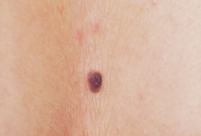 Figure X-1: Slightly raised compound nevus with regular borders and pigment | Photo courtesy of WebMD Opens in new window
Compound nevus is generally a well-circumscribed, small (<6mm) raised papule which is mostly uniformly pigmented with a range of color from skin-colored to tan to brown with either a rough or smooth surface. In this nevus, a proliferation of typical melanocytes involves both the dermoepidermal junction and the dermis.
Clinical Features
Clinically, compound nevus is characterized by the appearance of melanocytes not only at the junction but also in the dermis where they are organized in roundish nests, syncytia or lines of cells (cords or strands).
In a compound nevus the junctional component progressively fades away and in old lesions it is represented only by few nests and single cells.
Compound nevi are elevated relative to the surrounding uninvolved skin. They are often lighter in color than junctional nevi, but those that have been recently irritated may show areas of dark pigmentation.
Variants
Compound nevi have three major patterns:
- #1
The first one is flat with its lateral diameter being larger than the vertical one. The epidermis gently papillated and the intradermal component is restricted into the expanded papillary dermis or the superficial reticular dermis.
Such features as bridging of rete ridges, fibroplasias of the papillary dermis, lymphocytic infiltrates and a minor proportion of melanocytes with large nuclei are variably present, reflecting the biologic diversity of this entity and the possibility of a crisp separation between Clark and dysplastic nevus on histologic grounds.
- Unna nevus
The second kind of nevus, called Unna nevus, is characteristically exophytic with a markedly papillated silhouette.
The epidermis has an epidermal nevus or seborrheic keratosis – like pattern with elongated rete ridges which give a labyrinthine pattern by merging together.
Melanocytes are present in the core of the papillae. The dermal component of the nevus is restricted in the papillary dermis, which is greatly expanded; at the bottom a sharp longitudinal border demarcates the interface between the papillary and reticular dermis.
While Unna nevi are often clipped off with scissors, those that excised might have melanocytes around pilosebaceous units. In Unna nevi a special stain for elastic fibers frequently reveals a tight network of thick elastic fibers which resembles a nevus elasticus (probably a hint of the congenital nature of at least some of the Unna nevi).
- Miescher Nevus
Miescher nevus is characterized by the prevalent involvement of reticular dermis by the melanocytic proliferation.
This nevus has a domed surface and a wedge shape. The pigment is scarce and restricted to the superficial portion of the lesion or it is absent altogether.
Collagen fibers interposed among the cells are thick and coarse. Lateral and deep borders of the nevus in the reticular dermis are indistinct. Usually, the nevus has a very scanty junctional component which can be represented only by a lentiginous proliferation of single melanocytes without nests.
Histological Features
Compound nevi contain melanocytes Opens in new window in both the epidermis and dermis. The proliferation of melanocytes in the junctional component in compound nevi is the same as seen in junctional nevi; thus, melanocytes are distributed either in nests or single cells.
The junctional component usually does not extend at the lateral edges beyond the nested component; however, some nevi with lentiginous pattern (i.e., single cells along the rete ridges) may extend beyond the nested component.
The intradermal component in the papillary and reticular dermis has nests and cords of melanocytes, sometimes arranged in a coalescent, band-like pattern.
Nonetheless, these nests are equidistant rather than forming expansile nodules; thus, when large and confluent nests are seen in the dermis, this finding should raise suspicion for melanoma, similarly if nests at the deep aspect of the lesion are larger than the ones superficially.
The majority of acquired compound nevi do not extend beyond the papillary reticular dermis junction. The exception is nevi on the head and neck, since they commonly extend around hair follicles and in reticular dermis mimicking the pattern seen in congenital nevi Opens in new window.
There is a change in morphology with depth; the melanocytes in superficial dermis show a similar morphology as those seen in the junctional component, i.e., epithelioid cells with round nuclei and abundant cytoplasm with variable pigmentation; single nucleoli can be seen in the center of the nuclei (type A melanocytes).
The melanocytes deeper in the lesion are smaller and with minimal cytoplasm (type B melanocytes) or can be spindle with schwannian differentiation (type C melanocytes).
The stroma in the dermis is composed of collagen fibers arranged among single melanocytes and small nests. Large, irregular, and confluent sheets of melanocytes in the dermis without the presence of interposed collagen in between them are not a characteristic feature of a common nevus and should raise suspicion for melanoma.
Most nevi show maturation as characterized by the presence of progressive reduction of the size of melanocytes as well as by the decrease in size of the intradermal nests in the deeper aspects of the lesion. Also, the cells adopt spindle cell morphology and there is decreased pigmentation at the base of the lesion.