Hori's Nevus
Definition and Clinical Features
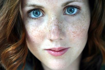 Figure X-1 | Photo courtesy of DR. Luciano Schiazza Opens in new window
Hori’s nevus, also known as acquired, bilateral nevus of Ota-like macules (ABNOM), is an acquired dermal melanocytosis characterized by multiple speckled blue-brown and/or slate-gray macules occurring bilaterally on the malar regions.
Less commonly, the temple, nose, forehead, and eyelids are affected. The appearance is very similar to that of nevus of Ota, but it does not extend to ocular or mucosal surfaces as in nevus of Ota Opens in new window.
Early lesions in Hori’s nevus consists of discrete border and tend to occur in clusters of blue-brown macules, whereas confluent slate-gray macules become more common over time.
Hori’s nevus is common in Asian women and usually presents after the third decade of life. Predisposing factors include chronic sun exposure, pregnancy, hormonal medication, and genetic influence.
Hori’s nevus can be distinguished clinically from nevus of Ota Opens in new window by its speckled and bilateral distributon, late onset in childhood, and lack of mucosal involvement.
Histologically, in ABNOM, elongated spindle-shaped melanocytes are dispersed between the collagen fibers and perivascularly in the upper dermis.
In contrast, melanocytes Opens in new window in nevus of Ota are distributed diffusely throughout the papillary and reticular dermis.
Therapeutic Considerations
- Pigment lesions lasers
The Q-switched ruby, the Q-switched alexandrite, and the Q-switched 1064 nm Nd:YAG lasers have all been used successfully for the treatment of Hori’s nevus. The degree of lightening is proportional to the number of treatments performed: the more treatments given, the better the results.
Typical parameters for the treatment of Hori’s nevus using the Q-switched ruby laser are 2–5 mm spot and fluence of 7–10 J/cm2; for the Q-switched alexandrite laser, 3 mm spot, 7.5–8 J/cm2; and for the Q-switched 1064 nm Nd:YAG laser, 3 mm spot, 4.2–9.9 J/cm2 (mean 7.8 J/cm2). The treatment endpoint was immediate whitening and treatment intervals range from two weeks to six months.
Hori’s nevus is known to be more resistant to laser treatment, requiring more sessions compared to nevus of Ota Opens in new window. One of the reasons for this is the higher rate of PIH seen after laser treatments for Hori’s nevus.
The incidence of PIH Opens in new window ranged from 7–73% of cases and was transient fading in 2–6 months within 4% hydroquinone cream.
The high rate of PIH Opens in new window may be related to the greater clustering of melanocytes around the blood vessels, as indirect vessel injury from the laser treatment leads to a higher degree of crythema and subsequently PIH.
Post-inflammatory hypopigmentation Opens in new window, which may be permanent, is more commonly seen with the shorter wavelength ruby and alexandrite lasers and also if the sessions are not adequately spaced.
- Topical agents
In Hori’s nevus, there is significant epidermal pigmentation. Methods to remove this epidermal pigmentation, thereby allowing more dermal penetration and greater efficacy, have been advocated.
The use of topical bleaching agents like hydroquinone and tretinoin to discharge epidermal melanin 4–6 weeks prior to Q-switched ruby laser was effective in Hori’s nevus with a low rate of PIH and decreased the number of laser sessions.
- Miscellaneous
Other combination treatments include the use of the scanned C02 laser to remove the epidermis followed by the Q-switched ruby laser has been shown to be more effective than Q-switched ruby laser alone in the treatment of Hori’s nevus.
Ee et al. showed that the combination of Q-switched 532 nm Nd: YAG laser (to remove epidermal hyperpigmentation) followed by the Q-switched 1064 nm Nd:YAG (532 nm, 4 mm spot, 1.2 J/cm2; and 1064 nm, 4 mm spot, 6.5 J/cm2) was more effective than Q-switched 1064 nm Nd:YAG laser alone for Hori’s nevus.
See also:
- Hori Y, Kawashima M, Oohara K, et al. (1984) Acquired, bilateral nevus of Ota-like macules. J AM Acad Dermatol 10:961-964.
- Ee HL, Wong HC, Goh CL, et al. (2006) Characteristics of Hori naevus: a prospective analysis. Br J Dermatol 154:50-53.
- Park JM. Tsao H, Tsao S. (2009) Acquired bilateral nevus of Ota-like macules (Hori nevus): etiologic and therapeutic considerations. J Am Acad Dermatol 61:88-93.
- Lee B, Kim YC, Kang WH, et al. (2004) Comparison of characteristics of acquired bilateral nevus of Ota-like macules and nevus of Ota according to therapeutic outcome. J Korean Med Sci 19:554-559.
- Kunachak S, Leelaudomlipi P, Sirikulchayanonta V. (1999) Q-switched ruby laser therapy of acquired bilateral nevus of Ota-like macules. Dermatol Surg 25: 938-941.
- Lam AYM, Wong DSY, Lam LK, et al. (2001) A retrospective study on the efficacy and complications of Q-switched alexandrite laser in the treatment of acquired bilateral nevus of Ota-like macules. Dermatol Surg 27:937-942.
- Polnikorn N. Tanrattanakorn S, Goldberg DJ. (2000) Treatment of Hori’s nevus with the Q-switched Nd:YAG laser. Dermatol Surg 26: 477-480.
- Suh DH, Han KH, Chung JH. (2001) Clinical use of the Q-switched Nd:YAG laser for the treatment of acquired bilateral nevus of Ota-like macules (ABNOMs) in Koreans. J Dermatol Treat 12:163-166.
- Momosawa A, Yoshimura K, Uchida G, et al. (2003) Combined therapy using Q-switched ruby laser and bleaching treatment with tretinoin and hydroquinone for acquired dermal melanocytosis. Dermatol Surg 29:1001-1007.